Panosteitis
A simplified way to describe panosteitis, sometimes referred to as pano, is growing pains. It occurs in young large or giant breed dogs, and is characterized by an acute onset of lameness with no associated trauma.
Typical age of onset is from 5-18 months but has been seen as young as 2 months and as old as 5 years. Males are affected four times more often than females. In females, the onset is often associated with their first heat. The cause is unknown, but with the high incidence in certain breeds (e.g., German Shepherd Dogs), a poly-genetic origin is suspected. Other causes may include stress, transient vascular abnormalities, metabolic disorders, allergies, hyperestrogenism, or autoimmune reactions following viral infection.
This disease usually presents itself with sudden onset of lameness. The lameness may be intermittent and varies in intensity, but rarely is the dog completely non-weight bearing. A unique characteristic is that there is often a shifting leg lameness, where it may resolve in one leg and then develop in another. Panosteitis is a disease of the shaft, not the joints, of the long bones. The most commonly affected bones, in order of frequency, are ulna, radius, humerus, femur, and tibia. The front limbs are usually affected first. In severe cases there may be a low grade fever, depression, reduced appetite, muscle loss or weight loss.
Suspicion of panosteitis arises when pain is elicited on firm palpation of the shaft of a long bone. Blood work is usually normal. Other conditions that need to be ruled out include hip dysplasia, osteochondritis dissecans, hypertrophic osteodystrophy, ununited anconeal process, fracture, ligamentous injury, immune mediated arthritides, and Lyme disease.
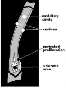
Radiographs will confirm the diagnosis. On x-rays of a lesion there will be areas of increased density (brighter white) and accentuated or loss of trabecular pattern (matrix of normal fibrous connective tissue) within the medullary cavity (core of shaft). The bone cortices (outer part of shaft) may be thickened. There may be smooth, linear periosteal proliferation (extra bone growth at the outermost covering of shaft). At later stages sclerotic (increased density or brighter white) areas will begin to decrease in size and density. Radiographic signs may persist months after lameness has resolved
The good news is that this is a self-limiting disease, but it may persist or shift for 6-18 months. Buffered aspirin or carprofen (Rimadyl) may alleviate some of the pain. Exercise restriction may be recommended for severe cases.
Sandra Wickwire, DVM
References
- The 5 Minute Veterinary Consult, Tilley & Smith, 1997, pp 904-905
- Radiographic Interpretation for the Small Animal Clinician, Owens, 1982, pp 16-17
- Saunders Manual of Small Animal Practice, Birchard & Sherding, 1994, pp 1073-75
This information is provided for educational purposes only and is not a substitute for medical advice. It is not to be used for diagnosing or treating a health problem, nor is it a substitute for professional care. If you suspect that your dog may have a health problem, please consult your veterinarian.